Saturday, June 4, 2016

my last poject

Assalamualaikum and hye. today i would like to share with you guys about my adopted  microbe which it is given name as Enterococcus faecalis which is as my last project for my microbe class


along the way to get know about my microbe characteristics, I just know that this microbe is very harmful to human and can cause death. from the name itself, we can know its shape is round shape. they are also part of the normal intestinal flora of humans and animals. they also have been long recognized as important human pathogens and are becoming increasingly so. they also can cause clinical infections in human. This bacteria have both an intrinsic and acquired resistance to antibiotics, making them important nosocomial pathogens. They are also resistance to penicillin beta-lactamases, chloamphenicol and others.

to get know about this microbe, do visit my powtoon scrapbook which I include some of the characteristic in it. here's my link. https://www.youtube.com/watch?v=t1B3y0i_09g 
i've done my best to do my scrapbook using this app. there are so many types of apps that we can use to make our scrapbook. but I found that this app is quite interesting and i hope you enjoy watch my scrapbook and get the information about this microbe more.

after that, on Friday before our class session for this semester ends, we have our 3 Minute Thesis presentation that is handle in DKU of my faculty. the presentation is not that good for me because i get too nervous because the hall is big and I got too many butterfllies in my stomach. but overall I manage to deliver what I want to say in this 3MT presentation. I wish I could given more opportunity to do my 3MT presentation again. because this is the best and good preparation and practice for me to improve my speaking and my delivery skill to people about my findings.
My 3MT slide

last but not least, I wish all the best to ALL of my classmate, CELL & MOLECULAR BIOLOGY students for our final exam that is just around the coner. my wishes all the best to you and answer the exam especially this microbiology course.

principles of ecology

in this chapter, i learnt about the habitat and all the population of each species. each microorganism are all depends on each others and most microbes also depend on themselves. in this chapter, we also learnt about the interaction among the microorganisms. there are two types of interactions which are positive and negative interaction.

the positive interaction are mutualism, cooperation and commensalism. while the negative interaction are predation, parasitism, amensalism and competition.

mutualism
predation
this kind of interaction is important for the ecosystem balance and the food chain can be regulate in a balance way.
our ecology system need to be preserved so that it can be balanced and many habitat can be saved for these animals to live

Thursday, May 19, 2016

trip to Institute of BioScience, UPM

this trip to ibs is on 25th March which we are going to have a look on different type of microscope that is being use by the scientist to determine and observed the microbe that they are going to have for their research. this ibs also is a place where research and development are being handle for the researchers to do their project and to observe on the microbe that they are looking for.

this is some of the pictures of the microbes that is being observed under transmission electron microscope
this are the types of  microscope that is being used in the lab


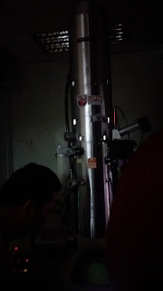
this is the electronic microscope that must be used under dark condition to observed the microbes.


meanwhile, this is a machine that is used where the sample is plated with gold.


this is our tour guide during the trip. where he is explaining all the type of microscope and machine that are in the IBS.


while walking through this IBS alley, I saw this cryogenic accessories that are used by them when doing their experiment for their protection towards the chemical that they might use during the experiment is being handle.


Wednesday, May 18, 2016

trip to yakult factory in seremban

we are having a trip to yakult factory on 16th May 2016 around 8 am. Our trip to yakult factory gives me a lot of new information about the bacteria that had been used on the production of yakult product. 

yakult is a world pioneer in the field of probiotics in the form of cultured milk drink. Born from the researcher of a doctor in 1935. yakult contain the probiotics bacteria called "Lactobacillus casei Shirota" also known as "Shirota strain". this cultured milk drink contain over 30 billion live Shirota strain in each bottlewhich they have the highest concentrations of probiotics compared to other probiotic products. The Shirota strain is scientifically to be the among of the strongest strains of beneficial bacteria and does give benefit to human health. Probiotic also known as kind of good bacteria. 

this bacteria lives in intestine which could make the system inner stronger. According to the Japanese Ministry of Health has approved that the Lactobacillus casei Shirota strain reaches the intestine alive, so this product maintains good health by increasing bacteria, decreasing harmful bacteria and improving the intestinal environment. Every different strain has different effect.

what I had learnt from this trip is that i get to know that:

why they develope yakult? because:
  • preventive medicine
  • a healthy intestine leads to a long life
  • healthy life for all with affordable price
the characteristics of this Shirota strain is that:
  • can reach our intestine alive
  • they resistant to artificial gastric juice in stomach which most of this bacteria still alive and can survive
  • they also resistant to bile juice
the beneficial of this bacteria is for:
  • health maintenance: 
    1. suppress the growth of harmful bacteria
    2. help digestion and absorption
    3. enhance immunity and resistance
the harmful bacteria caused bloated stomach and can cause diarrhea
factors:
  • balance of the intestinal flora is due:
    1. unbalanced diet
    2. stress
    3. aging
    4. infection
    5. antibiotics (medication)
the health benefit when consuming this cultured milk is that:
  • it can increase the good bacteria and reduce the harmful bacteria.
  • it can reduce the production of toxin in our body
  • it improve bowel movement
  • it boost in our immune system
this bacteria also active in temperature 15 to 45 degree celcius. 

this trip gives me an opportunity to get know deeper about this cultured milk drink and its benefits to our intestine.


Sunday, March 27, 2016

game based learning on eukaryote topic

For eukaryote topic we are having our own creative ideas to make the topic more interesting.


So we are having our own game based learning created by 6 groups. 

The first group presenting their game based on topic of cilia & flagella, cytoplasm and cell membrane. The game we are playing is a type of quiz question answer on Kahoot online application. This Kahoot app is very fun and interesting because it seems like we are playing games with its background music. All of us are excited to answer the questions quickly because it is given only 30 seconds to answer it. Therefore, it also help us to think faster within the time limit and got the correct answer at the same time.


Then we are playing crossword puzzle under topic golgi apparatus, centrosomes and chloroplast. This crossword puzzle is very fun because the questions that we are asked is quite difficult and confusing to answer it and we need to match the word with the boxes provided.


The third group are based on mitosis and meiosis topic which we are playing bingo where we need to complete 5 boxes in row to win. We need to fill up the answer provided into the 25 boxes then tick the right answer based on question given by them until a bingo is finish. This game required creativity and luck.

Next we are playing flip card which is under topic lysosome, vacuole and peroxisomes. In this game, each group need to answer the question to get the mark and we need to flip a pair of card based on the answer we give to get extra mark. Here, all of us are very excited because if a group didn’t get the correct card it will open the opportunity to flip another card for another group and given the marks. Game are always being played to get the marks.

lysosome structure


After that we are playing the game which we need to be as fast as we can to get the correct answer. The game is running with organelle under topic ribosome, nucleus and mitochondria. We need to be fast to get the answer because there is a lot small pieces of paper on the floor and we need to open them and find the correct answer based on the questions.


Last but not least, we are playing musical chair under topic smooth and rough endoplasmic reticulum. Each group will playing by two person and the person who didn’t get to sit on the chair need to answer the questions.

This kind of game based learning are very useful to make a productive, active and creative student to make the class more fun and interesting. I would like to have this learning game again.

That’s all from me. Thank you.
todays also our last class with Dr. Wan. so here we are. 

prokaryote the small microbes~


Today’s topic is on prokaryotic cells.


Eventhough I have learnt this topic since my secondary school then go on in my matriculation studies, I still learnt it now. I’m telling you it’s not going to be boring as long as you are still want to figure out and still want to know about it more and the fact is, I still can’t remember the name of the microbes that I’ve learnt before.

What have I learnt about prokaryotic is that they are very tiny microorganism and they are also simple compare to eukaryote. As everyone very familiar with these microorganisms, these prokaryote are divided into two categories which is the bacteria and archae. Both of them are similar but they are different. Bacteria can be differentiate with it cell wall either they lack of peptidoglycan or not and one of them have bigger periplasmic space which is the gram negative bacteria which the space contain hydrolytic enzyme.
Here are the cell wall structure for gram positive bacteria that i would like to share

this the cell wall structure of gram negative bacteria:
as you can see from above picture, the differences that they have is the gram negative does no contain techoic acid and it has thin layer of peptidoglycan compared to gram positive bacteria.

They also have these kind of external structure of cell wall which is glycocalyx, flagella, axial filament and pili. The structure that I thing I’ve never heard before is the glycocalyx. Glycocalyx is a coating that is secreted by the bacteria which is they use it to protect them from dehydration. It also contribute to bacterial virulence to which it can cause disease. they also known as capsule when it is organized and firmly attached to cell wall. when it is unorganized and loosely attached it is called slime layer.


They also have the structures internal to cell wall which include the plasma membrane, cytoplasm, nuclear area, ribosomes and inclusion. All of them are very familiar to me and all of my colleague. So I would like to share about the inclusion part which I’m not so familiar with it.

This inclusions are very important to the bacteria which is it act and serve as a basis of identification. The types of inclusions are:
·         Metachromatic granules
Which known collectively as volutin. It can be found in algae, fungi and protozoa. It contain polyphosphate for synthesis of ATP.  Example of algae that can be found is the Corynebacterium diphtheria.
Metachromatic granules

·        -  Polysaccharide granules
Consist glycogen and starch

·         - Lipid inclusions for lipid storage. It is stain by the Sudan dye

·         - Sulphur granules
It present in sulphur bacteria (thiobacillus) which they use sulphur as energy reserve.

·        -  Carboxysome
Contain enzyme ribulose 1,5-diphosphate carboxylase which is used for carbon fixation. They can be found in photosynthetic and nitrifying bacteria.

·        - Gas vacuoles
Found in aquatic prokaryotes in order to maintain buoyancy so that the cell can remain at the desire depth in the water.

·         Magnetosome
It act like a magnet to decomposed hydrogen peroxide which protect the cell from hydrogen peroxide accumulation.

Bacteria also produce endospore when they are in unfavourable conditions. This is because the endospore is highly resistant to environmental stress such as:
-         -  High temperatures
-         -  Irradiation
-          - Strong acid
-         -  Disinfectants


What I’m hoping from this topic is that I hope that I can be more interested to study about this microbe world because for me I’m not that good in remembering it really well about their characteristic with its functions because I might find it very difficult. So that I hope I can have more time to do some search on this this prokaryote topic.

Thursday, March 10, 2016

microscopy techniques


Assalamualaikum wbt.
Based on the picture above we all know that I’m going to share to you about microscope. Microscope is used worldwide by human this era.
Lets begin!

This topic lecture is very interesting and I think not everyone knows about the multifunction of each microscope that we have nowadays. learning on this topic makes me realize that even the small living things like bacteria and viruses can be seen and we can observed their internal structure as well. we can even see a 3D structure of the microorganisms under the specific microscope and we can see the exact shape of the microorganisms.. therefore we can know how do they look like and we can observe their motility under the microscope.


there are many types of microscope that I had learnt on this topic. the first one is the common use microscope which is the optical microscope and the electron microscope. 
The types of optical microscope are:


-dark field microscope


-bright field microscope

-fluorescence microscopy


-phase contrast microscopy:


The types of electron microscopy:

-scanning electron microscope:


-transmission electron microscope:


-environmental scanning electron microscope

each type of the microscope give different slide of specimen which is being observed and it helps the scientist to do their researched much better. the uses of microscope is very important to the scientists in order to observed the living things that can't be seen by our naked eyes especially small microorganisms such as bacteria and viruses.

microscope are also varies based on their magnification power where the resolution and refractive index are both important. the shorter the wavelength, the greater the resolution. other than that, under high magnification, the objective lens must be small and need to use oil immersion which has the same RI glass. in order to improve the resolution power.

there are many things that i learnt. what i am hoping from this lecture is that, i hope that I can differentiate and recognize the differences of the images on the specimens under each microscope. i also hope that i will find an alternative way to improve my learning skills on this topic. 

in this week also, we had a brief lecture on prokaryote. before we enter the lecture, Dr Wan assigned us to do some mind mapping  on this prokaryote topic using the applications that she provide to us so that we can understand more in the lecture. this is my mind mapping link that is very simple and good to refresh back what I had learnt.

this is my link for my mind map. have a nice brief to have a look for it. ;)

Sunday, March 6, 2016

Introduction of MICROBIOLOGY (Topic 1)


Assalamualaikum. For this semester I’m taking Microbiology subject. What I’m learning in this course is ALL about bacteria. I think that this course will be fun and interesting because as we all know our life is always surrounded by microorganism that called bacteria and others. In topic 1 for this course is the introduction about microbiology. What I learn in this topic is microbe is a living things that are too small to be seen by our naked eyes. So in order to see and observe them, we need to use microscope which O will learn about microscope in topic 2. Microorganism are also group into 5 categories which is bacteria, fungi, virus, protozoa and algae. We also learn about their characteristics and what are the differences between them that make them different function and benefits with each other. We also learn about how do they divide and their benefits in our lives.

Here are the examples of the microorganism that we had learnt in class.

E.coli bacteria

Protozoa

Algae bloom

Fungus

Lets the history begin!!
History is very important in order for us to know who found out the bacteria and what and how they do the experiment to observe the bacteria. The first observation is by Robert Hooke who is a most famous in his microscopial observation which is his study on thin slice of cork where he describe the microscopic world in his book called “Micrographia”. He also mark the beginning of Cell Theory which is all living things are composed of cell. Then the second scientist is Van Leeuwenhoek who proposed the “Spontaneous Generation”. This theory is proved by John Needham where he done his experiment that agreed with Anton theory. Then Lazzaro disapproved Needham’s theory. Rudolph Virchow then give a new concept called “BIOGENESIS” and says that “Living cells can arise only from its preexisting living cell. Then Pasteur was finally resolved the issue on concept of Spontaneous generation with Biogenesis by doing his own experiment.

this is the Pasteur's experiment where he disapprove the spontaneous generation theory that is proposed by Van Leeuwenhoek based on the theory that I found it is very interesting and important in the cell history
That is what I’ve learn in topic 1. ;)